Category Archives: News

MMSDG Présentation annuelle des étudiants et postdoc / Annual Trainees night

Suite à la présentation annuelle des étudiants et postdoc, voici les récipiendaires des prix des meilleurs présentations orale et par affiche :
————–
Following the annual trainees night, here is the winner for best presentation and best poster:

Best oral presentation :

Heath Patterson


Heath
Patterson – Ph.D. student
Department of Chemistry, Université de Montréal

Title: Phospholipid MALDI imaging mass spectrometry stratification of colorectal cancer liver metastasis clinical biopsies


Best poster presentation :

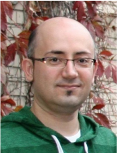
Makan Golizeh

Makan Golizeh – Ph.D. student
Department of Chemistry, 
Université du Québec à Montréal

Title: Identification of Liver Proteins Targeted by Reactive Metabolites using LC-MS/MS


Félicitation! Congratulation!

December 2, 2014 – John A. McLean

John McLeanDr John A. McLean

Professor of Chemistry at Vanderbilt University, Nashville, Tennessee, USA

“Targeting the untargeted: Structural mass spectrometry for the analysis of complex samples in systems, synthetic, and chemical biology”

Abstract:

One of the predominant challenges in systems-wide analyses is the broad-scale characterization of the molecular inventory in cells, tissues, and biological fluids. Advances in computational systems biology rely heavily on the experimental capacity to make panomics measurements, i.e. integrated metabolomics, proteomics, lipidomics, glycomics, etc., accompanied with fast minimal sample preparation, fast measurements, high concentration dynamic range, low limits of detection, and high selectivity. This confluence of figures-of-merit place demanding challenges on analytical platforms for such analyses. Ion mobility-mass spectrometry (IM-MS) provides rapid (ms) gas-phase electrophoretic separations on the basis of molecular structure and is well suited for integration with rapid (us) mass spectrometry detection techniques. Furthermore, the timescales of this multi-dimensional separation are well suited for combination with fast condensed-phase separations such as GC, SFC, and UPLC (min) for enhanced separation selectivity as the sample complexity becomes ever more challenging. This report will describe recent advances in IM-MS panomics measurement strategies in the analyses of complex biological samples of interest in systems, synthetic, and chemical biology. New advances in bioinformatics and biostatistics will also be described to approach biological queries from an unbiased and untargeted perspective and to quickly mine the data gathered to provide targeted and actionable information.

Brief Biography:

John A. McLean is Stevenson Professor of Chemistry at Vanderbilt University. Prof. McLean completed his Ph.D at George Washington University in 2001, where he made significant contributions in plasma spectrochemistry in the development of new technologies for the analysis of complex and limited radionuclide and biological samples. Subsequently, he performed postdoctoral research at Forschungszentrum Jülich in Germany with Prof. J. Sabine Becker and then as a postdoctoral at Texas A&M University with Prof. David H. Russell in biological mass spectrometry. Working with David Russell from 2001-2006, he constructed ion mobility-mass spectrometers capable of broad-scale analyses of extremely complex biological samples, termed ‘panomics,’ on the basis of both molecular structure and mass. In 2006, Prof. McLean was recruited to Vanderbilt University as Assistant Professor of Chemistry through both the Department of Chemistry and the Vanderbilt Institute of Chemical Biology. At Vanderbilt, McLean and colleagues focus on the conceptualization, design, and construction of structural mass spectrometers, specifically targeting complex samples in systems, synthetic, and chemical biology as well as nanotechnology. His group applies these strategies to forefront translational research areas in drug discovery, personalized medicine, and ‘human-on-chip’ synthetic biology platforms. Prof. McLean has received a number of awards, including the Agilent Thought Leader Award, Excellence in Teaching Award from the student members of the American Chemical Society, a Defense Threat Reduction Agency Research Award, an American Society for Mass Spectrometry Research Award, a Spectroscopy Society of Pittsburgh Award, an R&D 100 Award, and the Bunsen–Kirchhoff Prize from the GDCh (German Chemical Society), among others.

November 17, 2014 – Tommy Nilsson

Tommy NilssonDr Tommy Nilsson

Professor of Medicine and Director of Proteomics at McGill University

«New Cell Biology through the study of Human Fatty Liver Disease»

Human pathophysiology presents unique opportunities to uncover new molecular mechanisms. Here, human liver tissue derived from donors and patients were subjected to subcellular fractionation to enrich for organelles including lipid droplets, mitochondria, endoplasmic reticulum and the Golgi apparatus. Each fraction was then analyzed by liquid chromatography tandem mass spectrometry and matched against the human-centric database NeXtProt. Spectral counting was used to compare fractions in a semi-quantitative manner and following hierarchal clustering, compiled using Java TreeView to yield heat map-based distributions of each protein and protein neighborhood. Lipid droplet proteins previously never assigned to this organelle were uncovered and are presently being elucidated functionally. Liver-derived fractions were also subjected to lipid analysis through shotgun mass spectrometry and using ion intensity, compiled as above enabling correlation between proteins and lipid profiles. The value of studying human samples in the context of disease to generate new cell biology will be demonstrated through findings made.

October 14, 2014 – André Leblanc

AndreLeblancAndré Leblanc

Université du Québec à Montréal, roup of Lekha Sleno

2014 recipient of the Michel Bertrand award

Novel mass spectrometry-based methodologies to detect, characterize and quantify reactive drug metabolites and their protein adducts

Reactive metabolites are electrophilic and reactive species that can be metabolically generated from some xenobiotics. They have often been linked to toxicity via their ability to covalently bind to protein. Reactive metabolites generated from therapeutic drugs are responsible for a multitude of adverse drug reactions and impede the development of new drugs. The development of mass spectrometry-based analytical methods to study these species is needed to better understand the molecular processes involved with these types of toxicities. The talk will present several new methodologies pertaining to the detection and characterization of reactive metabolites and their covalent adducts.

May 13, 2014 – Bruno Domon

Bruno Domon

Bruno Domon

Centre de protéomique clinique, CRP-Santé, Luxembourg

Development of Proteomic Clinical Assays Based on High-Resolution / Accurate Mass Spectrometry

Targeted proteomics experiments applied to biomarker evaluation are routinely performed on triple quadrupole mass spectrometers in selected reaction monitoring (SRM) mode.  However, the low resolution of quadrupole mass filters has limited selectivity for complex samples, such as bodily fluids, characterized by a high biochemical background.  Hybrid mass spectrometers with high resolution and accurate mass (HR/AM) capabilities overcome this limitation, and have opened new avenues in quantitative proteomics.  The analyses of clinical samples using parallel reaction monitoring (PRM) on a quadrupole-orbitrap mass spectrometer showed significant gain in selectivity, while increasing the confidence in the measurements by identifying the fragments by accurate mass.

Furthermore, immuno-affinity isolation of proteins combined with a fast LC-MS technique has allowed the detailed characterization peptide isoforms of diagnostic cancer markers.  More specifically, the approach was applied to quantitatively profile EGFR and RAS protein isoforms, which both are predictive markers for targeted therapy.

April 16, 2014 – Christoph H. Borchers

Christoph H. Borchers

Christoph H. Borchers

Full Professor, University of Victoria, Director, UVic Genome BC Proteomics Centre

Novel Approaches in Quantitative and Structural Proteomics for Clinical Research and Diagnostics

Protein quantitation is essential for screening biomarker candidates for disease stratification and monitoring, and to verify and validate these biomarkers.  Accurate plasma protein concentrations can be determined through a targeted, multiplexed approach involving Multiple Reaction Monitoring (MRM), in conjunction with stable isotope-labeled standard (SIS) peptides. To improve the robustness of MRMs toward the analysis of thousands of patient samples, we have developed assays using standard-flow UPLC/MRM-MS for quantitating multiple proteins (>100) in undepleted human specimen including plasma, urine and CSF.  Recent developments in the modern mass spectrometry of proteins and peptides have resulted in significant progress in structural proteomics techniques for studying protein structure.  A variety of protein structural questions, ranging from defining protein interaction networks to the study of conformational changes and the structure of single proteins, can be addressed using multiple mass spectrometry based structural proteomics approaches.  Each technique provides specific structural information which can be used as experimental structural constraints in protein-structure modeling.  Here, we describe recent developments in limited proteolysis, surface modification, hydrogen-deuterium exchange, ion mobility, and crosslinking — all combined with modern mass spectrometric techniques — for studying the structure of clinical relevant proteins like prion.

Dr. Borchers received his B.S., M.S. and Ph.D. from the University of Konstanz, Germany.  After his post-doctoral training and employment as a staff scientist at NIEHS/NIH/RTP, in North Carolina, he became the director of the UNC-Duke Proteomics Facility and held a faculty position at the UNC Medical School in Chapel Hill, NC (2001-2006).  Since then, Dr. Borchers has been employed at the University of Victoria (UVic), Canada and holds the current positions of Professor in the Department of Biochemistry and Microbiology and the Don and Eleanor Rix BC Leadership Chair in Biomedical and Environmental Proteomics.  He is also the Director of the UVic – Genome BC Proteomics Centre, which is one out of five Genome Canada funded Science & Technology Innovation Centres and the only one devoted to proteomics.

His research is centred around the improvement, development and application of proteomics technologies with a major focus on techniques for quantitative targeted proteomics for clinical diagnostics.  Multiplexed LC-MRM-MS approaches and the immuno-MALDI (iMALDI) technique are of particular interest.  Another focus of his research is on technology development and application of the combined approach of protein chemistry and mass spectrometry for structural proteomics.  Dr. Borchers has published over 180 peer-reviewed papers in scientific journals and is the founder and CSO of two companies, Creative Molecules. Inc. and MRM Proteomics Inc.  He is also involved in promoting proteomic research and education through his function as HUPO International Council Member, Scientific Director of the BC Proteomics Network and Vice-President, External of the Canadian National Proteomics Network.

MMSDG (160414)

March 18, 2014 – Patrick Hayes

Patrick HayesPatrick Hayes

Professor, Analytical spectroscopy & environmental analyses, Chemistry department, U de M

Atmospheric Aerosol Composition and Sources in Los Angeles Studied by In-Situ Mass Spectrometry

Atmospheric aerosols have a well-known impact on human health and climate as highlighted in the most recent Intergovernmental Panel on Climate Change report published in 2013. However, atmospheric aerosols remain poorly characterized because, in part, of their chemical complexity. Specifically, the organic fraction of aerosols is composed of thousands of compounds that have a wide range of sources in the environment (e.g. diesel combustion and cooking emissions). This seminar will describe mass spectrometry measurements of organic aerosols carried out near Los Angeles as part of the CalNex field campaign. Five distinct chemical classes of organic aerosol are identified, and using the measured elemental compositions, mass spectra, and correlations with tracers each class can be associated with specific sources. Important findings include the observation that the total organic aerosol mass concentration is dominated by “secondary” organic aerosols, which are composed of the low volatility oxidation products of gaseous emissions.


Bio-sketch:

Patrick Hayes received his B.A. at Oberlin College in Ohio, and subsequently earned his Ph.D. in chemistry at Northwestern University under the guidance of Professor Franz Geiger. Dr. Hayes’ doctoral research investigated the interactions of adsorbates with geochemical solid/liquid interfaces using nonlinear optics. After completing his doctoral work, Dr. Hayes was a CIRES Postdoctoral Fellow at the University of Colorado with Prof. Jose-Luis Jimenez, and performed field measurements and modeling studies of aerosols in the Los Angeles metropolitan region. Dr. Hayes started as an assistant professor of analytical/environmental chemistry at Université de Montréal in August 2013.

MMSDG (180314)

February 18, 2014 – Benoit Coulombe

benoit_coulombe
Benoit
Coulombe

Director, gene transcription and proteomics laboratoyy at IRCM and professor investigator at the biochemistry depy. U de M

Interrogating regulatory mechanisms of protein complex assembly using functional proteomics

Our laboratory seeks to understand the mechanisms by which abnormal protein folding and protein complex assembly by molecular chaperones participate in the onset and the development of disease. We have discovered many proteins that regulate the function of molecular chaperones, including regulatory factors of nuclear RNA polymerase biogenesis and novel lysine methyltransferases that preferentially target and regulate molecular chaperones. Our work on chaperone methylation led us to propose the existence of a chaperone posttranslational modification code, termed the ‘‘chaperone code’’, which is at play to orchestrate the proper folding and assembly of protein complexes that make up the human proteome.

Benoit Coulombe is the director of the Gene Transcription and Proteomics Laboratory at the Institut de recherches cliniques de Montréal (IRCM) and a Professor of Biochemistry at the Université de Montréal.  He was an undergraduate in Biochemistry and obtained his Ph. D. in Molecular Biology at the Université de Montréal.  He undertook postdoctoral work at the University of Toronto under Jack Greenblatt and the Université libre de Bruxelles (ULB) under Albert Goldbeter (School of Ilya Prigogine, Nobel laureate in Chemistry).  He moved at the Université de Sherbrooke as an Assistant Professor in 1993 where he attained the tenured rank of Full Professor, and to the IRCM in Montréal in 2001.  Over the past 20 years he has explored the mechanisms by which regulated protein-protein, protein-DNA and protein-RNA interactions control the activity of RNA polymerase II during the transcription reaction.  His “Promoter Wrapping Model” for transcriptional initiation by multi-subunit RNA polymerases has been included in molecular biology textbooks.  Recently, Dr. Coulombe developed a proteomic procedure that his laboratory exploits to map and systematically monitor in patients protein interaction networks underlying neuromuscular and cardiometabolic diseases, thereby contributing to accelerate biomarker discovery and personalized drug treatment.  He is the director of the Canada Foundation for Innovation (CFI)-funded, Canada-wide National Technology Platform for Mapping Protein Interaction Networks in Health and Disease.  Dr. Coulombe was recently awarded the Bell-Bombardier Research Chair by the IRCM for his pioneering work in proteomics.

MMSDG (180214)

December 10, 2013 – Sponsors’ night

Image1

·        Jean-Francois Larouche, Phenomenex – Rapid Improvements for LC/MS/MS Analysis without Additional Method Development Using Phree™ phospholipids removal solutions.

·        Patrice Lemire, Waters – Multidimensional approach to Increased System Peak Capacity

·        Brigitte Simons, AB/Sciex – Advances in Front End MS Analytical Separations; Ion Mobility and Capillary Electrophoresis

·        Jean-Francois Roy, Agilent – A new high resolution Ion Mobility Quadrupole Time-of-Flight mass spectrometer for structural analyses

·        Bernard Delangh  , Thermo Fisher – An automated tool to create curated spectral libraries

November 12, 2013 – Terry D. Cyr

TerryCyrTerry Cyr

Senior scientist, Centre for Vaccine Evaluation, Health Canada, Ottawa, Ontario

Application of Mass spectrometry in the Regulation of Influenza Vaccines

Current methods for quality control of inactivated influenza vaccines include determining the amount of hemagglutinin (HA), verifying neuraminidase (NA) enzymatic activity, and demonstrating that the levels of the contaminant protein ovalbumin are below a limit. The HA assays require the availability of strain-specific reference HA antigens and antibodies. We have been developing rapid alternative methods for vaccine analysis that provide HA, NA and contaminant-protein identification and quantitation.  Enzymatically digested vaccine proteins are analyzed by LC–MSE and absolute quantification of the HA and NA antigens, other structural influenza proteins and chicken egg proteins is achieved by comparing the average intensity of their three most intense tryptic peptides (“Hi-3”) to those from a spiked reference protein.  In pursuing the validation of this method, we have expressed a protein that contains “Hi-3” peptide sequences from proteins of interest (QCONCAT) as well as compared our results to other MS and antibody based methods.  The results are wonderfully consistent, but not easily explained.

October 15, 2013 – Christina Bell

ChristinaBell

Christina Bell

University of Montreal

Molecularcharacterization of the contribution of autophagy to antigen presentation using quantitative proteomics

Autophagy, is a fundamental cellular degradation pathway that emerged as a new immunological paradigm of increasing interest. The application of quantitative proteomics methods allowed us to identify changes occurring during the remodeling of autophagosomes in response to disease and inflammatory conditions such as cytokine stimulation and viral infections. Bioanalytical mass spectrometry has played a key role in identifying and quantifying important regulators of autophagy in macrophages. Furthermore, our systems biology approach that combined mass spectrometry-based quantitative proteomics with shRNA screens revealed new biological insights on the molecular mechanisms governing the functions of the autophagosome in antigen presentation.

Prix Michel Bertrand en Spectrométrie de masse

This award was established earlier this year by the MMSDG to commemorate the pioneering work of Michel Bertrand in the field of bioanalytical Mass Spectrometry. This award is given to a graduate student from a Quebec University in his/her last year of doctoral studies and who made an exceptional contribution to the fundamental understanding and/or the application of mass spectrometry.

May 14, 2013 – Demian Ifa

Demian IfaDemian Ifa

Assistant professor, Chemistry department, York University

Mass Spectrometry Imaging under Ambient Conditions

Mass spectrometry imaging (MSI) has emerged as an important tool in the last decade and it is beginning to show potential to provide new information in many fields owing to its unique ability to acquire molecularly specific images and to provide multiplexed information, without the need for labeling or staining. In MSI, the chemical identity of molecules present on a surface is investigated as a function of their spatial distribution. In addition to now standard methods involving MSI in vacuum, recently developed ambient ionization techniques allow MSI to be performed under atmospheric pressure on untreated samples outside the mass spectrometer. In desorption electrospray ionization (DESI), a spray of charged droplets is directed at the sample creating a thin film of solvent on the surface. Further spray droplets collide with the solvent film and splash secondary droplets containing dissolved analytes into the air and from there they are directed into the mass spectrometer. DESI-MS imaging has become increasingly attractive due to its simplicity and the reduced sample preparation steps compared with vacuum based imaging techniques. Recent advances and applications of MSI under ambient conditions will be presented.

March 21, 2013 – Lars Konermann

LarsKonermannLars Konermann

Professor and Canada Research Chair, Department of Chemistry University of Western Ontario

Analyzing Proteins by ESI Mass Spectrometry: Fundamentals and Biophysical Applications

Electrospray ionization (ESI) generates intact gas-phase ions from analytes in solution for a wide range of mass spectrometric investigations. Over the past few years our laboratory has contributed to the development of a comprehensive ESI mechanistic framework. The view that emerges from experimental and computational studies implies that ESI can proceed via three different avenues. Low molecular weight analytes follow the ion evaporation model (IEM), whereas the charged residue model (CRM) applies to large globular species. A chain ejection model (CEM) has been proposed for unfolded proteins and other disordered polymers. This presentation will also discuss recent ESI-MS applications, highlighting novel approaches for investigating protein folding and conformational dynamics. These techniques involve the use of rapid mixing techniques, hydrogen/deuterium exchange, as well as covalent labeling..

January 22, 2013 – Dajana Vuckovic

DajanaVuckovicShort bioDajana Vuckovic just joined Concordia University as an Assistant Professor in Bioanalytical Chemistry at the Department of Chemistry and Biochemistry. She holds Honours B.Sc. in Chemistry from the University of Toronto and Ph.D. in Analytical Chemistry from the University of Waterloo.  Her doctoral research under supervision of Prof. Janusz Pawliszyn focused on the development of in vivo solid-phase microextraction methodology for global metabolomics and small rodent pharmacokinetic studies.  As NSERC Postdoctoral Fellow at the University of Toronto with Prof. Andrew Emili, she developed novel chemical proteomics workflow for the determination of protein targets of drugs. She is the recipient of several awards including Johnson & Johnson Young Scientist Scholarship and 2010 Douglas E. Ryan Graduate Student Award by Canadian Society for Chemistry. She has co-authored 28 publications and 8 book chapters to date, and contributed 30 oral and 10 poster presentations at national and international conferences. She is currently the Editor of Sample Preparation, and an Editorial Board Member of Bioanalysis and Journal of Integrated Omics. At Concordia, she plans to establish a state-of-the-art research program in analytical and clinical metabolomics with particular focus on the development of new strategies to improve metabolome coverage of unstable and low abundance metabolites and the development of improved diagnostic methods for bipolar disorder.

Abstract :

The omission of metabolism quenching step in metabolomics studies of biological fluids can result in undesirable changes in metabolome composition and adversely impact subsequent data quality and interpretation.  In vivo solid-phase microextraction (SPME) is a new and effective sample preparation method for untargeted LC-MS metabolomics studies which incorporates metabolism quenching step directly during the sampling process and eliminates the need for blood withdrawal. In this study, the performance of in vivo SPME sampling of circulating mouse blood was compared against the traditional approaches based on blood withdrawal in combination with solvent precipitation, ultrafiltration and ex vivo SPME in order to study the effect of single carbamazepine dose on endogenous metabolism of mice. The overall metabolite coverage of SPME was lower than that of solvent precipitation and ultrafiltration when a single coating was used for extraction, but can be further enhanced with the use of complementary coatings. More importantly, in vivo SPME successfully captured unstable metabolites not observed by any of the traditional methods, with approximately 4% unique SPME features including important metabolites such as carotenes and β-NAD and improved quantitation of many unstable metabolites, for example glutathione or adenosine. Overall, we demonstrate that in vivo SPME can play an important role in untargeted studies to accurately capture metabolome at the time of sampling as well as in targeted studies of unstable and short-lived metabolites. The second part of the talk will focus on a novel chemical proteomics method for the determination of protein targets of bioactive molecules using chromatographic fractionation. The proposed method, called target identification by chromatographic co-elution (TICC), relies on the observable shift of the elution profile of a small ligand when the ligand is bound to a protein under non-denaturing chromatographic conditions. The methodology was validated using well-studied ligands with known interacting protein partners, such as interaction of trichostatin A with HDAC1 (Ki=3.4 nM) and sordarin with ELF2 (Kd = 1.26 µM). We also successfully identified ASC1 and DAK1 as novel targets of A77636 showing the capability of technique for de novo target deconvolution and its promise in both phenotype-based and target-centric discovery pipelines.

December 4, 2012 – Steve Carr

Steve Carr

Steve Carr

Director of Proteomics at the Broad Institute of MIT and Harvard

Quantitative Proteomics in Biology, Chemistry and Medicine

A new era of quantitative biology enabled by mass spectrometry based proteomic technologies has arrived. We can now define the content, relative abundance, modification states and interaction partners of proteins in a dynamic and temporal manner on a near-global basis in organelles, whole cells and clinical samples, providing information of unprecedented detail.  At the Broad Institute we are employing these technologies in a wide array of studies including delineating the genetic underpinnings of mitochondrial disorders, connecting cancer genotype to molecular phenotype, unraveling the basis of the innate-immune response, identifying the mechanism of action of drug-like molecules and to discover and verify protein biomarkers of disease. A representative set of project vignettes will be presented to convey a sense of the breadth and depth of application of modern proteomics to biology and medicine.

November 20, 2012 – Kevin Bateman

Kevin BatemanKevin Bateman

Leader Global Bioanalytical Scientific Network Senior Bioanalytical Scientist for PPDM and Director of the Methods Development group for Merck’s Regulated Bioanalytical Group

Recent Advances in Applied Mass Spectrometry in Drug Discovery

Over the past decade, we witnessed a significant transformation in the use of drug metabolism and pharmacokinetic (DMPK) data in support to the discovery and development of novel therapeutic compounds. Mass spectrometry has played a key role in supporting the lead optimization phase of drug discovery, and the recent introduction of high resolution mass analyzers and high-speed data processing systems further enhanced the value of MS-based technologies in metabolite detection and identification This presentation will highlight examples of the application of contemporary MS techniques to the optimization of drug candidates in an industrial drug discovery setting, and the complementary roles of high resolution MS in metabolite structure elucidation.

October 29, 2012 – Alain Brunelle

Alain BrunelleAlain Brunelle

Head of MS group, ICSN-CNRS, Gif-sur-Yvette, France

Biological applications of lipid imaging with cluster-TOF-SIMS and MALDI-TOF

This lecture intends to show the wealth of powerful information that can be obtained from the chemical analysis of biological surfaces, with several examples chosen among various applications such as the localization of xenobiotics, natural substances, lipid markers from genetic diseases and non-alcoholic fatty liver disease. The strengths and weaknesses of lipid imaging using TOF-SIMS and MALDI-TOF will also be compared, showing the complementarity between the two methods.